
Intrinsic Mitochondrial Apoptosis Pathway
Cellular stress-triggered Bax/Bak pore formation, Cytochrome c release, and Apaf-1 apoptosome assembly with Caspase-9 activation.
MOMP
Use the Apoptosis Diagram Generator to turn programmed cell death cascades into clear, journal-quality figures. Map intrinsic mitochondrial release, extrinsic Fas/TRAIL receptor signaling, apoptosome assembly, and executioner Caspase-3/7 cleavage.
Free to try ·
Your diagram will appear here
Describe what you need and click Generate
Turn programmed cell death cascades into clear, verifiable scientific figures ready for peer-reviewed journal submission.
Input your target apoptotic stimulus, death receptor ligands, Bcl-2 family members, apoptosome cofactors, and executioner substrates.
Check proteolytic activation arrows against direct inhibition bars (Bcl-2, XIAP) to ensure rigorous biochemical pathway logic.
Download layered SVG or PPTX presentation slides to adjust protein shapes, colors, and font sizes without resolution loss.
Input your target apoptotic stimulus, death receptor ligands, Bcl-2 family members, apoptosome cofactors, and executioner substrates.
Check proteolytic activation arrows against direct inhibition bars (Bcl-2, XIAP) to ensure rigorous biochemical pathway logic.
Download layered SVG or PPTX presentation slides to adjust protein shapes, colors, and font sizes without resolution loss.
Explore editable scientific diagrams detailing intrinsic mitochondrial permeabilization, extrinsic death receptors, caspase cascades, and cell death morphology.
Cellular stress-triggered Bax/Bak pore formation, Cytochrome c release, and Apaf-1 apoptosome assembly with Caspase-9 activation.
FasL and TRAIL receptor trimerization recruiting FADD to form the DISC complex and activate initiator Caspase-8.
Hierarchical proteolytic activation connecting initiator caspases to executioner Caspase-3/7 and cleavage of PARP and ICAD.
Molecular balance between pro-survival Bcl-2/Bcl-xL, BH3-only sensitizers (Bim, Puma), and pore-forming Bax/Bak effectors.
Cellular progression through chromatin condensation, nuclear fragmentation, membrane blebbing, and macrophage phagocytosis.
Bcl-2 and Beclin-1 regulatory switch balancing autophagic cell survival against irreversible apoptotic commitment.
SciDraw can help draft a scientific visual. Check all labels, relationships, and claims against your source before use.
Workflow Need | SciDraw AI | Generic Image AI |
|---|---|---|
Biochemically Rigorous Cleavage Logic | Automatically formats active cleaved caspase heterodimers distinct from inactive zymogens, with clear arrowheads and cleavage sites. | Blurs inactive procaspases with active cleaved subunits and places arrows inconsistently. |
Multi-Compartment Organelle Architecture | Renders crisp double-membrane mitochondria with intermembrane space, outer membrane pores, and nuclear lamina boundaries. | Draws floating proteins without double-membrane mitochondrial or nuclear boundaries. |
Full Vector & Presentation Slide Export | Export high-resolution SVGs and editable PPTX files with ungroupable protein labels and scalable vector shapes for journal submission. | Outputs flattened raster images where protein labels cannot be edited or ungrouped. |
Review Rights Before Use | Paid plans permit commercial use under the SciDraw License, subject to applicable law and third-party rights. Check your publisher’s disclosure requirements. | Unclear usage terms or unverified visual claims that complicate peer-review compliance. |
Follow proven scientific visualization principles to ensure your diagrams are accurate, legible, and aligned with journal requirements.
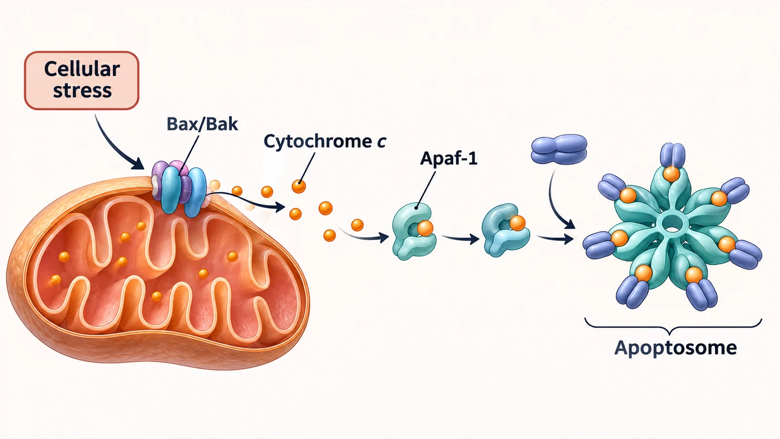
Apoptosis represents a tightly coordinated program of cell death essential for tissue homeostasis and development. Accurate scientific visualization requires maintaining clear separation between the two primary activation routes while highlighting points of crosstalk:
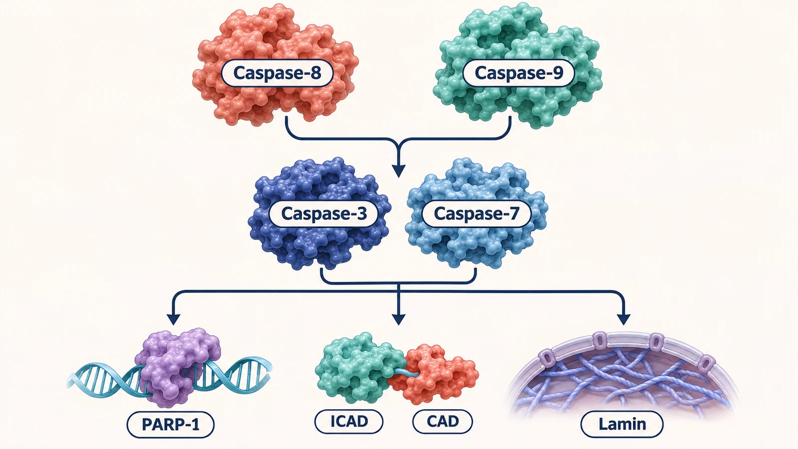
Peer-reviewed journals enforce strict visual standards for biochemical figures to avoid confusing experimental results with established pathways:
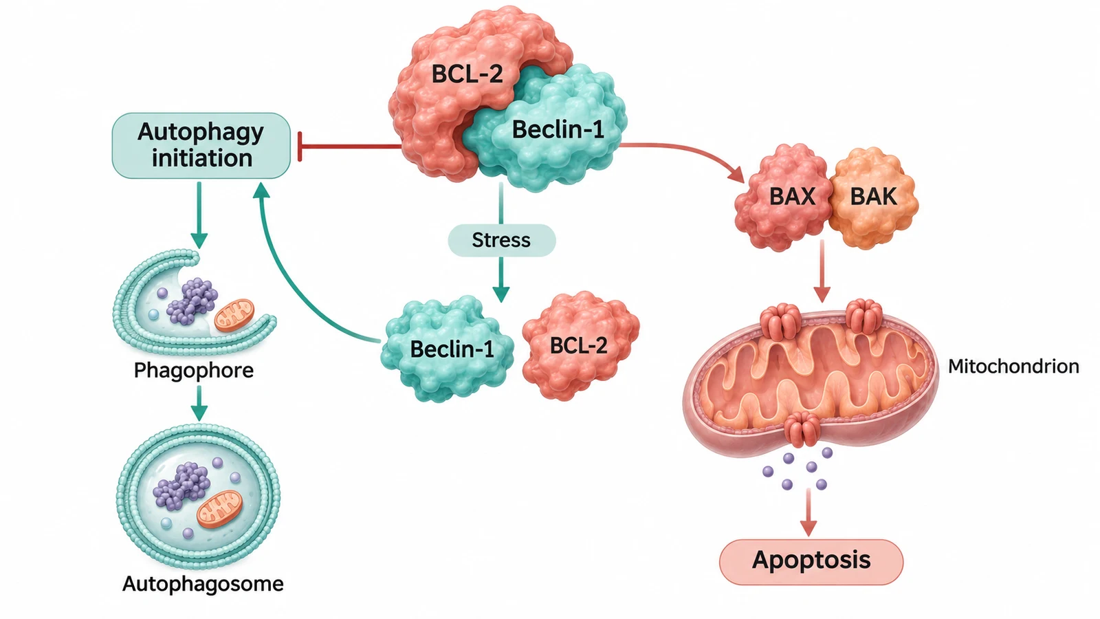
When submitting figures created with SciDraw AI to academic publishers, researchers should follow transparent attribution and experimental validation protocols:
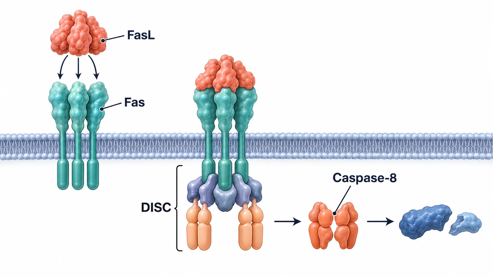
A single pathway or mechanism figure often serves multiple communication needs—from a two-column manuscript panel to a 16:9 conference slide or grant schematic. Plan your visual hierarchy so labels and compartments remain clear across formats:
Choose your plan. No hidden fees, cancel anytime.
Your data is yours · Never used to train any model · Encrypted in transit and at rest
Great for getting started
Includes
Best for regular creation
Includes
Built for high-volume production
Includes









Researchers, graduate students, and educators share how SciDraw helps them create scientific figures, scientific diagrams, graphical abstracts, and clearer teaching visuals faster.
For a plant-cell lecture, I needed the chloroplasts and vacuole called out without rebuilding an old slide. SciDraw gave me a useful scientific figure layout that went into my slide deck after only a few label edits.

Dr. Helen Vance
Cell Biology Professor
When a student asks for a force-diagram example beyond the textbook, I open SciDraw instead of redrawing one on a slide. It is the scientific diagram generator I reach for first, then I quickly check every vector.

Dr. Javier Ortega
Physics Instructor
For grant proposals, we use SciDraw to agree on the experimental setup diagram before anyone starts polishing the scientific figure. It has cut down the back-and-forth and given the team a shared visual from day one.

Dr. Devendra Verma
Research Lab Director
I can compare a few biological pathway diagrams for study notes before I settle on one.

Minh Nguyen
Graduate Science Student
For my dissertation, I start the manuscript-to-figure process with neural-pathway and synapse layouts in SciDraw. It gives me a strong first composition, so I can spend Illustrator time on the details that matter.

Dr. Ananya Rao
PhD Candidate · Neuroscience
The first scientific drawing got our fractional-distillation design review moving right away.

Dr. Marcus Davis
Chemical Engineering Lead
If the stock image does not match that week’s climate topic, I make a quick diagram in SciDraw. It gives the class a visual that fits the discussion, and I can tailor the labels to their level.

Prof. Mei-Ling Zhao
Environmental Science Educator
I see authors use a scientific figure generator to turn a rough outline into something reviewers can actually read. It makes the first review conversation clearer and helps the science stand out from the layout.

Dr. Thomas Ødegård
Scientific Journal Editor
For tectonics classes, I use a custom volcano cross-section diagram when the stock image does not match the boundary we are discussing. Students grasp the process much faster when the visual follows the exact example.

Dr. Fatima Al-Sayed
Geology Lecturer
It gives me a tidy scientific figure draft for a force diagram or wave sketch quickly, without making the curriculum page feel crowded.

Dr. Nathan Brooks
STEM Developer
I place a simple materials-science diagram next to SEM images so students can connect an ideal lattice to the real sample. That visual comparison makes the lesson click much faster than another paragraph of explanation.

Dr. Clara Sommer
Materials Researcher
Before opening CAD, we use SciDraw’s architecture diagram generator to make the system boundary and handoffs visible. It gets the right questions onto the table early and helps the team align before detailed modeling.

Dr. Robert Söderberg
Mechanical Engineer
For photosynthesis slides, I start with a chloroplast scientific figure that I can tailor to the level of the class. It saves a surprising amount of preparation time when a lesson needs a fresh visual.

Dr. Hannah Weber
Plant Biology Researcher
It gets me past the blank page on a science explainer and gives the final illustration a clearer direction.

Ji-Min Kim
Scientific Content Creator
Having students create a scientific drawing for a lab report makes them think through the process step by step. The discussion and revision afterwards are noticeably stronger.

Dr. Sofia Alvarez
Biochemistry Professor
It is helpful when we need an astronomy diagram for the exact orbit or instrument configuration in the discussion, rather than a generic solar-system graphic. The result feels much closer to the research question.

Dr. David Park
Astronomical Researcher
A labeled ecology figure gives our postdoc meetings something concrete to point at when we discuss feedback loops. It keeps the discussion focused and helps us reach a paper figure faster.

Dr. Lisa Wang
Ecology Postdoc
For multi-panel papers, I use the manuscript-to-figure workflow to decide what each panel needs to do before Illustrator. It makes the first layout decisions faster and leaves more time for the final polish.

Carlos Ortega
Illustrator Assistant
For early circuit and network reviews, I use SciDraw’s architecture diagram generator to get a first pass on the page. The team can mark it up together instead of building slides from fragments, which makes decisions quicker.

Dr. Elena Gomez
Systems Engineering Lead
For public-facing science guides, clear scientific diagrams often travel better than a dense block of text. They make complex ideas easier to share across languages and give us a strong starting point for the final guide.

Dr. Michael Achebe
Science Communicator
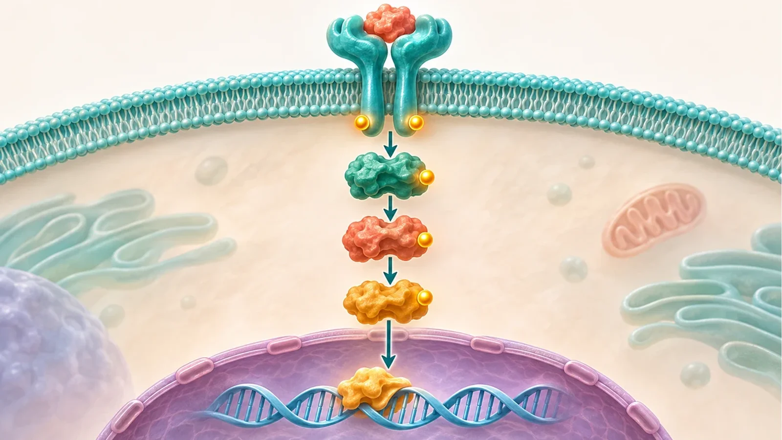 Research
Create diagrams for receptor cascades, phosphorylation relays, and transcriptional responses.
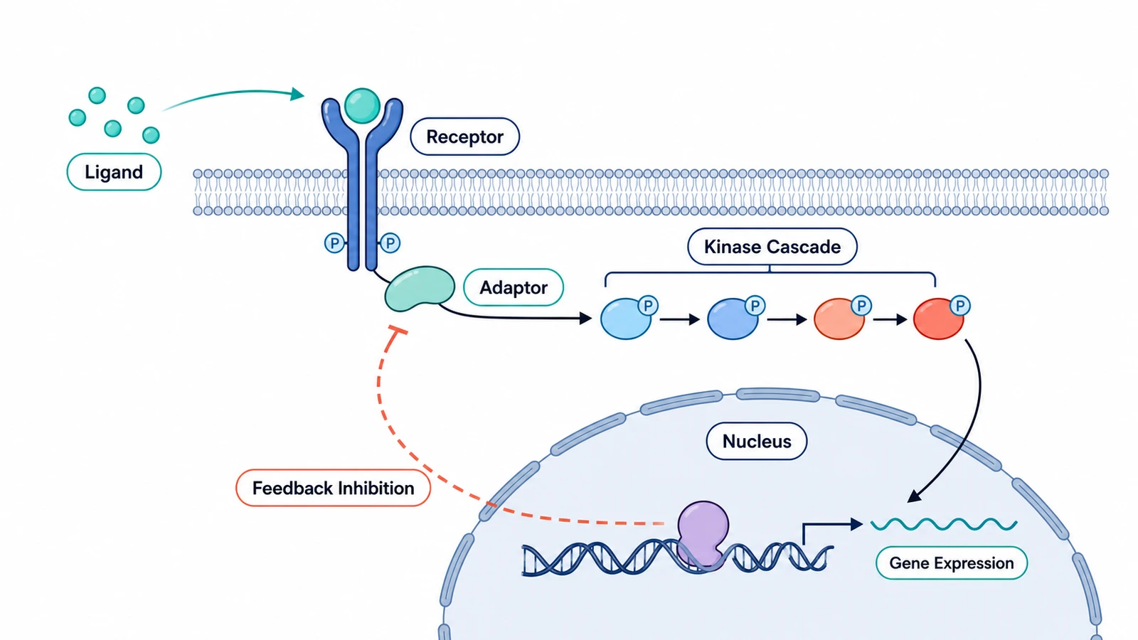 Research
Visualize biological cascades, metabolic networks, and molecular mechanisms.
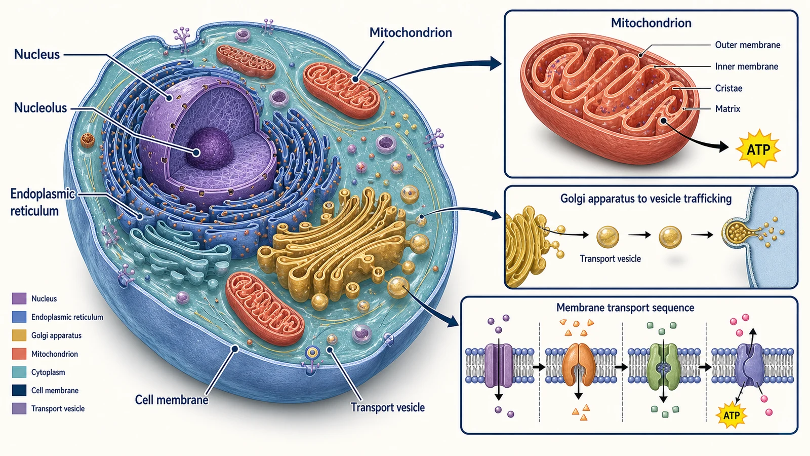 Research
Generate detailed eukaryotic and prokaryotic cellular cross-sections.